45 external structure of the heart with labels
Heart Anatomy: size, location, coverings and layers ... The heart wall is composed of three layers: the epicardium, myocardium, and endocardium. Location of the heart in the mediastinum. The superficial epicardium is the visceral layer of the serous pericardium. The middle layer is the myocardium and is composed mainly of cardiac muscle and forms the bulk of the heart. Heart Anatomy | Anatomy and Physiology - Lumen Learning The wall of the heart is composed of three layers of unequal thickness. From superficial to deep, these are the epicardium, the myocardium, and the endocardium. The outermost layer of the wall of the heart is also the innermost layer of the pericardium, the epicardium, or the visceral pericardium discussed earlier. Figure 6.
Human Heart (Anatomy): Diagram, Function, Chambers ... The heart is a muscular organ about the size of a fist, located just behind and slightly left of the breastbone. The heart pumps blood through the network of arteries and veins called the ...

External structure of the heart with labels
Chapter 22 Heart Flashcards | Quizlet Label the coronary arteries in an anterior view of the heart. Label the order that blood flows through in the heart, using the arrows as guides. Label the components of the heart wall. Label the components of the heart as seen from a posterior view. Label the major coronary veins. Label the components of the conduction system. Andrew File System Retirement - Technology at MSU Information technology resources, news, and service information at MSU. It is maintained by IT Services and the Office of the CIO. Anabolic steroid - Wikipedia Anabolic steroids, also known more properly as anabolic–androgenic steroids (AAS), are steroidal androgens that include natural androgens like testosterone as well as synthetic androgens that are structurally related and have similar effects to testosterone. They increase protein within cells, especially in skeletal muscles, and also have varying degrees of virilizing …
External structure of the heart with labels. The Anatomy of the Heart, Its Structures, and Functions The heart is the organ that helps supply blood and oxygen to all parts of the body. It is divided by a partition (or septum) into two halves. The halves are, in turn, divided into four chambers. The heart is situated within the chest cavity and surrounded by a fluid-filled sac called the pericardium. This amazing muscle produces electrical ... A Guide on the Structure of heart, its Chambers and valves The heart is a muscular organ situated in the middle of the chest slightly to the left.. It is a conical-shaped hollow organ enclosed within a pericardium, the peritoneal counterpart for the heart.. It is placed obliquely, behind the body of the sternum and costal cartilages, one-third of it is to the right, and the rest two-third lying left of the median plane. A Labeled Diagram of the Human Heart You Really Need to ... The human heart, comprises four chambers: right atrium, left atrium, right ventricle and left ventricle. The two upper chambers are called the left and the right atria, and the two lower chambers are known as the left and the right ventricles. The two atria and ventricles are separated from each other by a muscle wall called 'septum'. Heart Anatomy: Heart Dissection The letters indicated in the text refer to the labels on the picture. The anterior surface of the heart is characterized by the presence of the large arteries leaving the base of the heart, the pulmonary trunk (H) and the aorta (G). The pulmonary trunk is the vessel that divides to give rise to the two pulmonary arteries going to each lung.
How to Draw the Internal Structure of the Heart (with ... Once you have the basic outline of the heart sketched out, use an existing diagram to help you fill in the additional veins and muscles, like the mitral and aortic valves. After you've drawn the structure, color the different sections of the heart distinct colors and appropriately label them. Music industry - Wikipedia Labels outside of these three major labels are referred to as independent labels ... Business structure. The main branches of the music industry are the live music industry, the recording industry, and all the companies that train, support, supply and represent musicians. The recording industry produces three separate products: compositions (songs, pieces, lyrics), recordings … Lesson | The Heart - External Structure | Encounter Edu To be able to label a diagram of the external structure of the heart correctly identifying arteries and veins To be able to identify where blood enters and leaves the heart Expedition Prep Checklist Download the Google Expeditions App on all devices and select the expedition The Heart. The structure of the heart - Structure and function of the ... It is located in the middle of the chest and slightly towards the left. The heart is a large muscular pump and is divided into two halves - the right-hand side and the left-hand side. The...
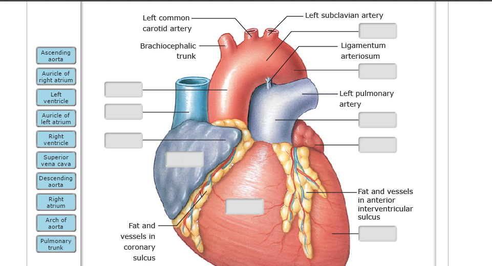
PDF Anatomy of Heart Labeled and Unlabeled Images (a) Anterior view of the external heart C' 2019 Pearson Education. Aort'c arch Ligamentum arteriosum Left pulmonary artery Left pulmonary ve ns Auricle of left atrium Circumflex artery Left coronary artery (in atrioventricular sulcus) Great cardiac vein Left ventricle Anterior interventricular artery (in anterior interventricular sulcus) Apex Human Heart - Diagram and Anatomy of the Heart Because the heart points to the left, about 2/3 of the heart's mass is found on the left side of the body and the other 1/3 is on the right. Anatomy of the Heart Pericardium. The heart sits within a fluid-filled cavity called the pericardial cavity. The walls and lining of the pericardial cavity are a special membrane known as the pericardium. Ch. 19 Circulatory System- heart Flashcards | Quizlet Correctly label the external anatomy of the anterior heart. Place the labels in order denoting the flow of blood through the pulmonary circuit beginning with the right atrium and ending in the left atrioventricular valve. The first and last structures are given. Right atrium 1. tricuspid valve 2. right ventricle 3. pulmonary valve How Strategy Shapes Structure - Harvard Business Review In this structuralist approach, structure shapes strategy. But as Kim and Mauborgne, the authors of Blue Ocean Strategy, point out, history reveals plenty of situations in which firms ...
Aqua Fanatic: 07/01/2011 - 08/01/2011
Structure of the Heart | SEER Training The outer layer of the heart wall is the epicardium, the middle layer is the myocardium, and the inner layer is the endocardium. Chambers of the Heart The internal cavity of the heart is divided into four chambers: Right atrium Right ventricle Left atrium Left ventricle The two atria are thin-walled chambers that receive blood from the veins.

The Heart | S-cool, the revision website
en.wikipedia.org › wiki › DapagliflozinDapagliflozin - Wikipedia Dapagliflozin, sold under the brand names Farxiga (US) and Forxiga (EU) among others, is a medication used to treat type 2 diabetes. It is also used to treat adults with certain kinds of heart failure and chronic kidney disease.
😀 Drag the labels to identify structural components of the heart. Label the heart — Science ...
Human Heart - Anatomy, Functions and Facts about Heart The heart wall is made up of 3 layers, namely: Epicardium - Epicardium is the outermost layer of the heart. It is composed of a thin-layered membrane that serves to lubricate and protect the outer section. Myocardium - This is a layer of muscle tissue and it constitutes the middle layer wall of the heart.

Europe - ThinEbook E-books
The Structure of Musical Preferences: A Five-Factor Model Much of the research concerned with music preferences has focused on questions pertaining to its structure and external correlates; very few studies have actually examined the contexts in which people listen to music and the particular music they listen to. As a result, most of the research in this area conceptualizes preferences as trait-like constructs and assume that …

Label the heart - Science Learning Hub In this interactive, you can label parts of the human heart. Drag and drop the text labels onto the boxes next to the diagram. Selecting or hovering over a box will highlight each area in the diagram. Pulmonary vein Right atrium Semilunar valve Left ventricle Vena cava Right ventricle Pulmonary artery Aorta Left atrium Download Exercise Tweet
Surfaces and Borders of the Heart - TeachMeAnatomy Sulci of the Heart. The heart is a hollow structure. On the interior, it is divided into four chambers. These divisions create grooves on the surface of the heart - these are known as sulci. The coronary sulcus (or atrioventricular groove) runs transversely around the heart - it represents the wall dividing the atria from the ventricles ...

Biology Diagrams,Images,Pictures of Human anatomy and physiology : Heart External Structure ...
Solved Art-Labeling Activity: Overview of the external ... Science; Anatomy and Physiology; Anatomy and Physiology questions and answers; Art-Labeling Activity: Overview of the external anatomy of the heart anterior view Res Great cardiac vein Aortic arch Right coronary artery Left coronary artery Left pulmonary veins Ascending aorta Left pulmonary artery Anterior interventricular artery Superior vena cava Pulmonary trunk Auricle of left atrium ...
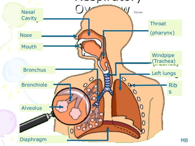
Human respiration
Label the Heart - The Biology Corner Shows a picture of a heart with letters and blanks for practice with labeling the parts of the heart and tracing the flow of blood within the heart.

The Science Scoop: Heart Diagram
Layers of the heart: Epicardium, myocardium, endocardium ... The heart is a muscular organ found in the middle mediastinum that pumps blood throughout the body. It is housed in the pericardial sac, which protects it and assists with its mechanics. Recalling from the heart anatomy, it has two atria and two ventricles that make up elements and important steps for the heart cycle.
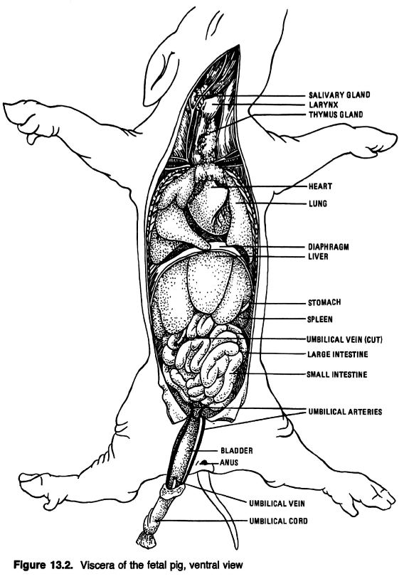
Anatomical Drawings of a Fetal Pig
The Heart - Science Quiz - Seterra The Heart - Science Quiz: Day after day, your heart beats about 100,000 times, pumping 2,000 gallons of blood through 60,000 miles of blood vessels. If one of your organs is working that hard, it makes sense to learn about how it functions! This science quiz game will help you identify the parts of the human heart with ease. Blood comes in through veins and exists via arteries—to control the ...
31 Label The Structures Of The Thoracic Cavity - Labels Database 2020
Internal Structure of the Heart | Contemporary Health Issues It is marked by the presence of four openings that allow blood to move from the atria into the ventricles and from the ventricles into the pulmonary trunk and aorta. Located in each of these openings between the atria and ventricles is a valve, a specialized structure that ensures one-way flow of blood.

the unlabelled structure of heart - Google Search | Heart diagram, Parts of the heart, Nursing ...
Solved Help Label the external anatomy on this posterior ... Science. Biology. Biology questions and answers. Help Label the external anatomy on this posterior view of a mammalian heart by clicking and dragging the labels to the correct location Coronary sinus Apex of heart Lert atrium Posterior interventricular branch of LCA Left pulmonary artery Left ventricle Left pulmonary veins Aortic arch.

Biology 156: April 2012
External anterior heart labeling Quiz - PurposeGames.com About this Quiz. This is an online quiz called External anterior heart labeling. There is a printable worksheet available for download here so you can take the quiz with pen and paper.
Heart Anatomy Cross Section Diagram Stock Vector 157286378 - Shutterstock
Heart - External Features - Anatomy QA Apex beat. Is the lowermost and outermost thrust of the heart, felt on the front of the chest. In adults it is felt in the left 5 th intercostal space 9cm. from the median plane (just medial to the midclavicular line). In infants it is felt in the 3 rd intercostal space just lateral to the midclavicular line.. Dextrocardia. It is a congenital anomaly in which the heart lies on the right side ...

Medical Encyclopedia - Structure: Structure of the Heart - Aviva | THE HEART | Pinterest | Heart ...
Structure Of The Heart | A-Level Biology Revision Notes The heart is a hollow muscular organ that lies in the middle of the chest cavity. It is enclosed in the pericardium, which protects the heart and facilitates its pumping action. The heart is divided into four chambers: The two atria (auricles): these are the upper two chambers. They have thin walls which receive blood from veins.

Heart and blood vessels - online presentation
Heart Anatomy Labeling Game This is an online quiz called Heart Anatomy Labeling Game. There is a printable worksheet available for download here so you can take the quiz with pen and paper. Your Skills & Rank. Total Points. 0. Get started! Today's Rank--0. Today 's Points. One of us! Game Points. 19. You need to get 100% to score the 19 points available.

Cardiovascular System - Anatomy And Physiology
Anabolic steroid - Wikipedia Anabolic steroids, also known more properly as anabolic–androgenic steroids (AAS), are steroidal androgens that include natural androgens like testosterone as well as synthetic androgens that are structurally related and have similar effects to testosterone. They increase protein within cells, especially in skeletal muscles, and also have varying degrees of virilizing …
Post a Comment for "45 external structure of the heart with labels"