45 images of compound microscope with labels
Compound microscope Images, Stock Photos & Vectors - Shutterstock 3,117 compound microscope stock photos, vectors, and illustrations are available royalty-free. See compound microscope stock video clips Image type Orientation Sort by Popular Science College and University Biology Insects and Spiders Jobs/Professions microscope laboratory compound eye optical microscope scientist Next of 32 Looking at the Structure of Cells in the Microscope A typical animal cell is 10–20 μm in diameter, which is about one-fifth the size of the smallest particle visible to the naked eye. It was not until good light microscopes became available in the early part of the nineteenth century that all plant and animal tissues were discovered to be aggregates of individual cells.
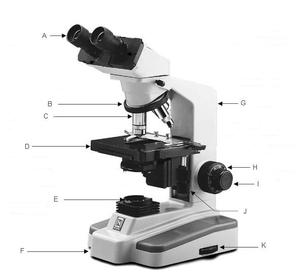
Parts of a Compound Microscope and Their Functions Eyepiece lens: It's a lens that's attached to the body tube's top structure of microscope.On the rim, a number (5X, 10X, 15X) is always written. This number represents the compound microscope magnification power of the device. With the eyepiece, you may see a magnified image of the thing.

Images of compound microscope with labels
16 Parts of a Compound Microscope: Diagrams and Video 4. Eyepiece. The eyepiece, also known as the "Ocular", is the first magnification lens you will look through in a compound microscope. Put simply, this is where you put your eye to see the image. Usually eyepieces come in 10X magnification, or 15X magnification but they can vary from 5X - 30X. Compound Microscope - Types, Parts, Diagram, Functions and Uses Image 5: The arm of the compound microscope. Picture Source: zfic.org Arm - it supports the head of the microscope and attach it to the base. Image 6: The revolving nosepiece. Picture Source: img-aws.ehowcdn.com Nosepiece - It holds the objective lens and attaches them to the head of the microscope. Compound Microscope Labeled Diagram - Quizlet QUESTION. The total magnification of a specimen being viewed with a 10X ocular lens and a 40X objective lens is. 15 answers. QUESTION. a mosquito beats its wings up and down 600 times per second, which you hear as a very annoying 600 Hz sound. if the air outside is 20 C, how far would a sound wave travel between wing beats. 2 answers.
Images of compound microscope with labels. (PDF) Introduction to Microscopy - ResearchGate compound microscope [4] 4. ... • TEM images are simply magnified images of the electron ... This new microscope doesn't require any special labels and could help increase access to low-cost ... Amazon.com: My First Lab Duo Scope Microscope - Young … KIDS MICROSCOPE KIT -- This kids microscope kit, in addition to the user manual and experiment guide, includes 5 blank slides, 1 concavity (well) slide, 4 prepared slides, 2 bottles of stain, 5 slide labels, 5 cover glasses, 50 sheets of lens paper, 1 plastic transfer pipette, 1 plain wooden applicator, 1 cotton-tipped applicator, 1 plastic forceps, 1 plastic test tube and 1 plastic … Compound Microscope Stock Illustrations - Dreamstime New users enjoy 60% OFF. 181,491,921 stock photos online. Download 711 Compound Microscope Stock Illustrations, Vectors & Clipart for FREE or amazingly low rates! New users enjoy 60% OFF. 181,491,921 stock photos online. ... Clearly labeled vector of modern compound microscope. EPS 8 with no gradients or effects, layers labeled for easy editing. Parts of the Microscope with Labeling (also Free Printouts) Parts of the Microscope with Labeling (also Free Printouts) A microscope is one of the invaluable tools in the laboratory setting. It is used to observe things that cannot be seen by the naked eye. Table of Contents 1. Eyepiece 2. Body tube/Head 3. Turret/Nose piece 4. Objective lenses 5. Knobs (fine and coarse) 6. Stage and stage clips 7. Aperture
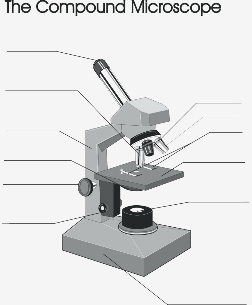
Electron microscope - Wikipedia An electron microscope is a microscope that uses a beam of accelerated electrons as a source of illumination. As the wavelength of an electron can be up to 100,000 times shorter than that of visible light photons , electron microscopes have a higher resolving power than light microscopes and can reveal the structure of smaller objects. Food Calorimetry: How to Measure Calories in Food We have the compound microscope you are looking for! Digital Microscopes. Digital microscopes are great for large classroom computer combined instruction. Students can take images, videos, and more. Stereomicroscopes. Stereomicroscopes show 3D images vs. flat images and are easier to focus and use. They are great for first tme student use. Compound Microscope Parts - Labeled Diagram and their Functions - Rs ... The term "compound" refers to the microscope having more than one lens. Basically, compound microscopes generate magnified images through an aligned pair of the objective lens and the ocular lens. In contrast, "simple microscopes" have only one convex lens and function more like glass magnifiers. [In this figure] Two "antique ... Parts of a Compound Microscope - Labeled (with diagrams) Image 1: The figure above is the standard image of a compound microscope. image source: 5.imimg.com The structural components of a compound microscope. Picture 2: The basic parts of a compound microscope. image source : optimaxonline.com Head/body it is where the upper optical parts of the microscope can be found. Base
Compound Microscope Stock Photos and Images - Alamy Compound Microscope Stock Photos and Images (1,984) Narrow your search: Vectors | Black & white | Cut Outs Page 1 of 20 A view down the eye piece of a upright compound microscope ID: DWCF6K (RF) Compound microscope, cut-out ID: BXR7RC (RM) Zeiss standard compound microscope ID: BH07H6 (RM) non illuminated compound microscope in a display exbibition Microscope Parts and Functions Microscope Parts and Functions With Labeled Diagram and Functions How does a Compound Microscope Work?. Before exploring microscope parts and functions, you should probably understand that the compound light microscope is more complicated than just a microscope with more than one lens.. First, the purpose of a microscope is to magnify a small object or to magnify the fine details of a larger ... Labeling the Parts of the Microscope | Microscope World Resources Labeling the Parts of the Microscope. This activity has been designed for use in homes and schools. Each microscope layout (both blank and the version with answers) are available as PDF downloads. You can view a more in-depth review of each part of the microscope here. Microscope Components - Science Quiz - GeoGuessr Microscope Components - Science Quiz: The most common type of modern microscope is called a compound microscope. They have two systems of lenses, one is the eyepiece and the other is comprised of one or more objective lenses. This type of microscope has become so advanced that some are capable of magnifying up to 1000 times! Microscopes are used in …
Compound microscope - YouTube
Compound Microscope with labels Stock Vector | Adobe Stock Download Compound Microscope with labels Stock Vector and explore similar vectors at Adobe Stock. Adobe Stock Photos Illustrations Vectors Videos Audio Templates Free Premium Editorial Fonts

Labeling a Compound Microscope Quiz
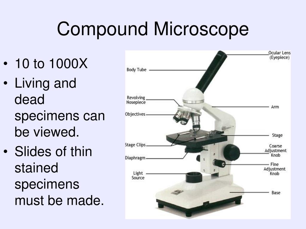
Compound Microscope Parts, Diagram Definition, Application, Working ... A compound microscope can magnify the image of a tiny object up to 1000. The term compound means "multiple" or "complex". The compound microscopes is consists of two lenses includes, the objective lens (typically 4x, 10x, 40x or 100x) in a rotating nosepiece closer to the specimen, and the eyepiece lens (typically 10x) in the binocular ...

Microscope With Labels Clip Art at Clker.com - vector clip art online, royalty free & public domain
Compound microscope - their parts and function - Microscopy4kids Compound microscopes generate magnified images through an aligned pair of the objective lens and the ocular lens. In contrast, "simple microscopes" have only one convex lens and function more like glass magnifiers. Eyepiece (ocular lens) The eyepiece (or ocular lens) is the lens at the top of a microscope that the viewer looks through. The ...
Parts of the Microscope Flashcards | Easy Notecards
Compound Microscope Parts, Functions, and Labeled Diagram Compound Microscope Definitions for Labels. Eyepiece (ocular lens) with or without Pointer: The part that is looked through at the top of the compound microscope. Eyepieces typically have a magnification between 5x & 30x. Monocular or Binocular Head: Structural support that holds & connects the eyepieces to the objective lenses.

Chapter 1 Questions PPT - BIOLOGY JUNCTION
Multiphoton Microscopy | Nikon’s MicroscopyU Two-photon excitation microscopy (also referred to as non-linear, multiphoton, or two-photon laser scanning microscopy) is an alternative to confocal and deconvolution microscopy that provides distinct advantages for three-dimensional imaging.In particular, two-photon excitation excels at imaging of living cells, especially within intact tissues such as brain slices, embryos, whole …

Microscope World Blog: Lactobacillus under the Microscope
Compound Microscope: Definition, Diagram, Parts, Uses, Working ... - BYJUS Compound microscope is a type of optical microscope that is used for obtaining a high-resolution image. There are more than two lenses in a compound microscope. Learn about the working principle, parts and uses of a compound microscope along with a labeled diagram here.
29 You Will Love Labeling A Compound Microscope — db-excel.com
Compound Microscope- Definition, Labeled Diagram, Principle, Parts, Uses The optical microscope often referred to as the light microscope, is a type of microscope that uses visible light and a system of lenses to magnify images of small subjects. There are two basic types of optical microscopes: Simple microscopes. Compound microscopes. The term "compound" in compound microscopes refers to the microscope having ...

Choosing a microscope « Adafruit Industries – Makers, hackers, artists, designers and engineers!
Parts of Stereo Microscope (Dissecting microscope) – labeled … The difference between Compound and Stereo (Dissecting) Microscope. Unlike a compound microscope that can only see a very thin specimen, stereo microscopes can be used for viewing almost anything you can fit under them. However, stereo microscopes offer lower magnification, typically 5x-50x, comparing to compound microscopes.
The Parts Of A Microscope Quiz - ProProfs Quiz
What is a Compound Microscope? - Microscope Clarity A compound microscope utilizes a system of compounding lenses that enables the microscope to produce highly magnified images. Some of the lenses involved in this compound lens structure are the condenser lens, objective lens (which are themselves made up of several lenses), and the eyepiece lens. Compound microscopes can produce images magnified anywhere from 40x - 2,500x.
PPT - cells PowerPoint Presentation, free download - ID:6853122
Label the image of a compound light microscope - Soetrust Which was the first cell viewed by the light microscope? Which of the following is true regarding the properties of… The compound below is treated with n-bromosuccinimide (nbs)…

label the parts of the compound microscope - Brainly.ph
Looking at the Structure of Cells in the Microscope A typical animal cell is 10–20 μm in diameter, which is about one-fifth the size of the smallest particle visible to the naked eye. It was not until good light microscopes became available in the early part of the nineteenth century that all plant and animal tissues were discovered to be aggregates of individual cells. This discovery, proposed as the cell doctrine by Schleiden and …

Microscope With Labels Clip Art at Clker.com - vector clip art online, royalty free & public domain
Cell Types Gizmo Worksheet - Name: Date: Student Exploration: … Gizmo Warm-up In the Cell Types Gizmo, you will use a light microscope to compare and contrast different samples. On the LANDSCAPE tab, click on the Elodea leaf. (Turn on Show all samples if you can’t find it.) Switch to the MICROSCOPE tab to observe the sample as it would appear under the microscope.

32 Label Of Compound Microscope - Label Design Ideas 2020
What is a Compound Microscope? - New York Microscope Company A compound microscope is an instrument that is used to view magnified images of small specimens on a glass slide. It can achieve higher levels of magnification than stereo or other low power microscopes and reduce chromatic aberration. It achieves this through the use of two or more lenses in the objective and the eyepiece.

PZ C: compound microscope
300+ Free Microscope & Laboratory Images - Pixabay Find your perfect microscope image. Free pictures to download and use in your next project. 189 37. analysis biochemistry. 335 71. analysis biochemistry. 334 96. microscope slide. 725 186.

Choosing a Microscope | Make: DIY Projects and Ideas for Makers
What is Electron Microscopy? - UMASS Medical School Conventional scanning electron microscopy depends on the emission of secondary electrons from the surface of a specimen. Because of its great depth of focus, a scanning electron microscope is the EM analog of a stereo light microscope. It provides detailed images of the surfaces of cells and whole organisms that are not possible by TEM.
Post a Comment for "45 images of compound microscope with labels"