44 microscope images with labels
Microscope Labeled Pictures, Images and Stock Photos Browse 49 microscope labeled stock photos and images available, or start a new search to explore more stock photos and images. Newest results Fluorescent Imaging immunofluorescence of cancer cells growing... Microscope diagram vector illustration. Labeled zoom instrument... Microscope diagram vector illustration. 22 Parts Of a Microscope With Their Function And Labeled Diagram Microscope Description A microscope is a laboratory instrument used to examine objects that are too small to be seen by the naked eye. In other words, it enlarges images of small objects. Invented by a Dutch spectacle maker in the late 16th century, light microscopes use lenses and light to magnify images. Generally a microscope ... Read more 22 Parts Of a Microscope With Their Function And ...
18,701 Microscope drawing Images, Stock Photos & Vectors - Shutterstock Microscope drawing royalty-free images 18,701 microscope drawing stock photos, vectors, and illustrations are available royalty-free. See microscope drawing stock video clips Image type Orientation Color People Artists Sort by Popular Science Abstract Designs and Shapes College and University Art Styles Printing, Typography, and Calligraphy

Microscope images with labels
Microscope Types (with labeled diagrams) and Functions The working principle of a simple microscope is that when a lens is held close to the eye, a virtual, magnified and erect image of a specimen is formed at the least possible distance from which a human eye can discern objects clearly. Simple microscope labeled diagram Simple microscope functions It is used in industrial applications like: 400+ Free Microscope & Bacteria Images - Pixabay 412 Free images of Microscope Related Images: bacteria laboratory science scientist research biology lab virus microscopic Find your perfect microscope image. Free pictures to download and use in your next project. Explanation and Labelled Images - New York Microscope Company The samples are labeled with fluorophore where they absorb the high-intensity light from the source and emit a lower energy light of longer wavelength. The resulting fluorescent light is then separated from the surrounding radiation with filters, allowing the observer to see only the fluorescing material.
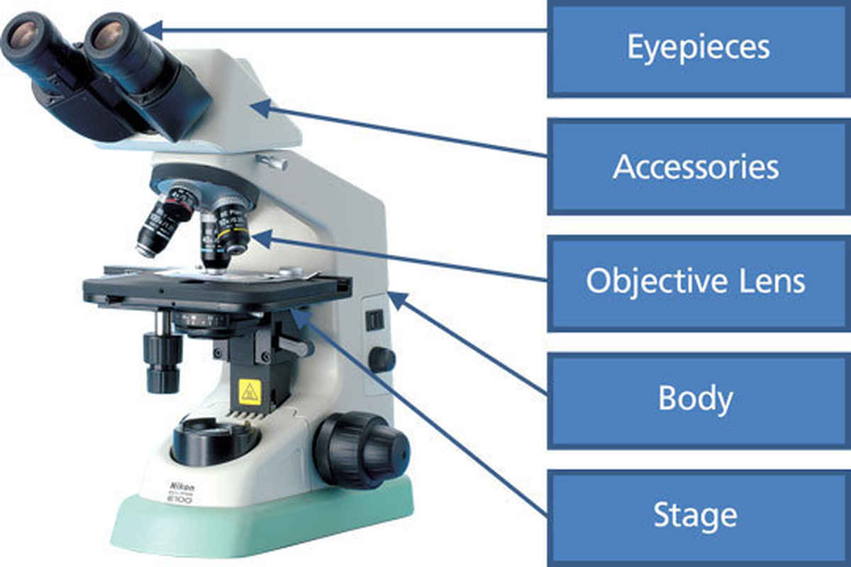
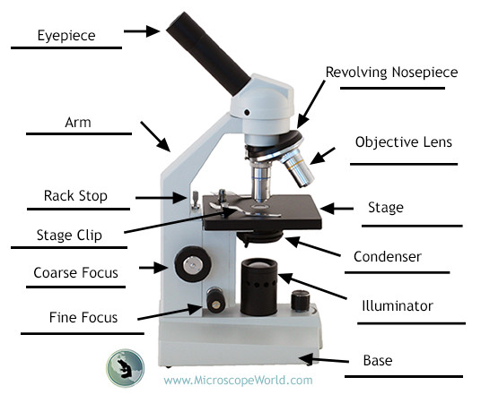
Microscope images with labels. Microscope Parts, Function, & Labeled Diagram - slidingmotion Microscope parts labeled diagram gives us all the information about its parts and their position in the microscope. Microscope Parts Labeled Diagram The principle of the Microscope gives you an exact reason to use it. It works on the 3 principles. Magnification Resolving Power Numerical Aperture. Parts of Microscope Head Base Arm Eyepiece Lens Microscope Parts and Functions First, the purpose of a microscope is to magnify a small object or to magnify the fine details of a larger object in order to examine minute specimens that cannot be seen by the naked eye. Here are the important compound microscope parts... Eyepiece: The lens the viewer looks through to see the specimen. A Study of the Microscope and its Functions With a Labeled Diagram ... The camera present within the microscope captures images to reveal the finer details of the specimen. This microscope can zoom and view the density of a specimen until it is only a micrometer thick and has a magnification ranging between 1,000 - 250,000x on the fluorescent screen. This microscope needs a computer software to yield precise ... Compound Microscope - Diagram (Parts labelled), Principle and Uses See: Labeled Diagram showing differences between compound and simple microscope parts Structural Components The three structural components include 1. Head This is the upper part of the microscope that houses the optical parts 2. Arm This part connects the head with the base and provides stability to the microscope.
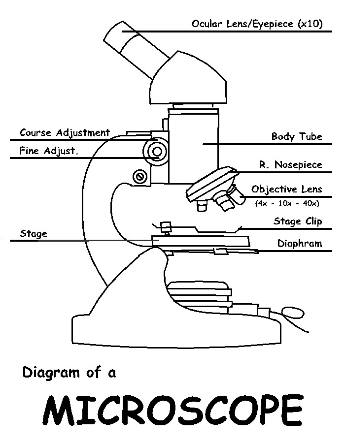
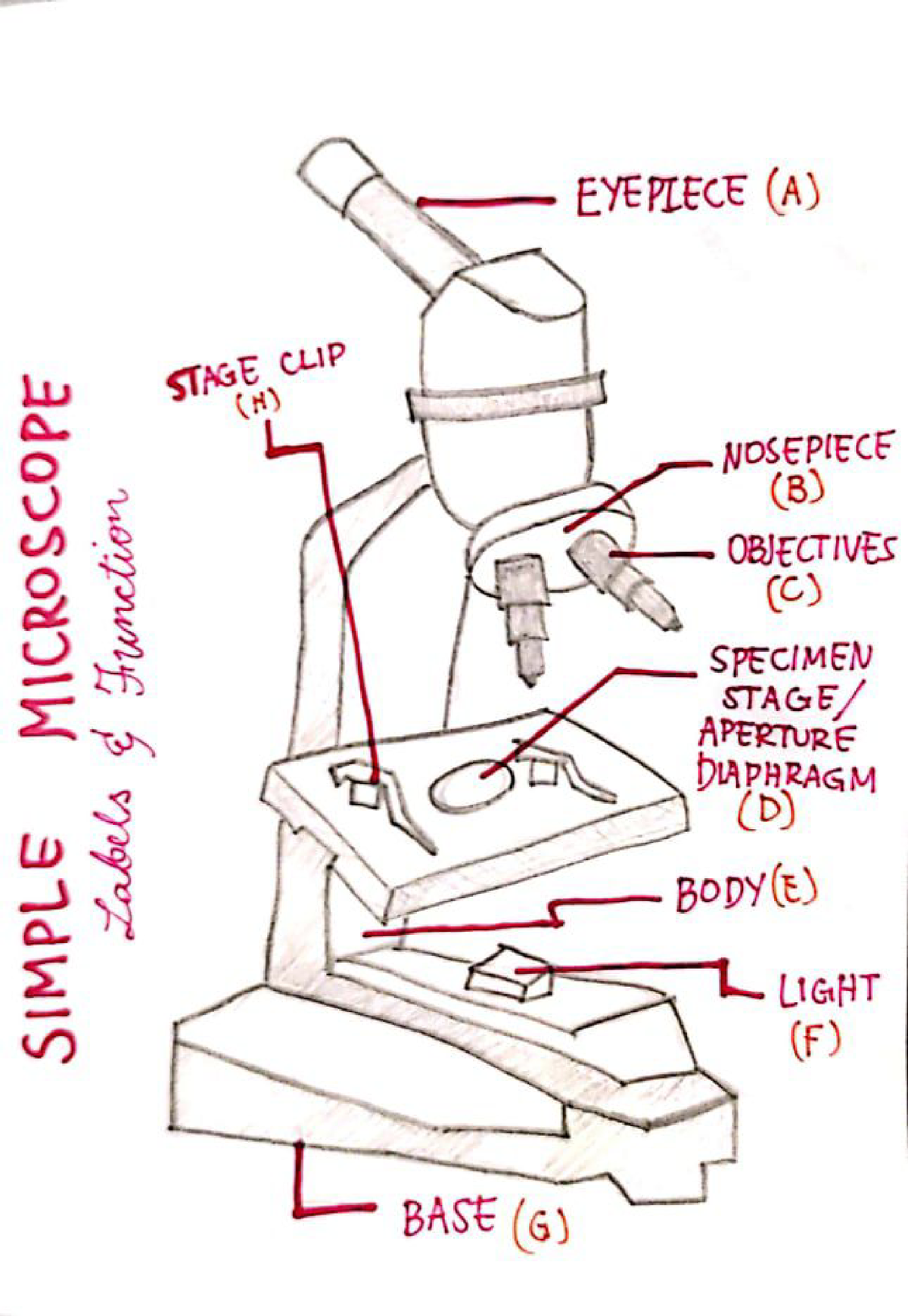
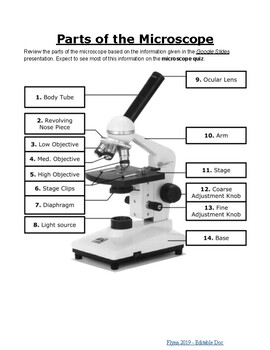
Simple Microscope - Parts, Functions, Diagram and Labelling Confocal microscope - It uses laser light to scan a dyed sample. Scanning electron microscope - Instead of light, this type of microscope uses electron. This type of microscope is used by researchers in the field of physical, biological, and medical science. Transmission electron microscope - it uses electron to create a magnified image ... Binocular Microscope Anatomy - Parts and Functions with a Labeled ... Now, I will discuss the details anatomy of the light compound microscope with the labeled diagram. Why it is called binocular: because it has two ocular lenses or an eyepiece on the head that attaches to the objective lens, this ocular lens magnifies the image produced by the objective lens. Binocular microscope parts and functions Compound Microscope Parts - Labeled Diagram and their Functions The eyepiece (or ocular lens) is the lens part at the top of a microscope that the viewer looks through. The standard eyepiece has a magnification of 10x. You may exchange with an optional eyepiece ranging from 5x - 30x. [In this figure] The structure inside an eyepiece. The current design of the eyepiece is no longer a single convex lens. Microscope Labeling - The Biology Corner The google slides shown below have the same microscope image with the labels for students to copy. I often spend the first day walking students through the steps and having them look at a single slide as we do the steps. Students are often very enthusiastic about using microscopes and will try to start with the high power objective.
473,241 Microscope Images, Stock Photos & Vectors | Shutterstock Microscope royalty-free images 473,689 microscope stock photos, vectors, and illustrations are available royalty-free. See microscope stock video clips Image type Orientation People Artists Sort by Popular Science College and University Healthcare and Medical Jobs/Professions Biology microscope laboratory scientist medicine research Next of 4,737 Polarizing Microscope Image Gallery | Science Lab - Leica Microsystems The position of the optical axis can be clearly determined with circular polarization. Right: Conoscopic image of the same calcite sample with linear polarized light. The calcite section is perpendicular to the optical axis. Images recorded with a DM4 P microscope using transmitted light, conoscopy, 63x N Plan objective, and polarizers. Parts of a microscope with functions and labeled diagram - Microbe Notes Parts of a microscope with functions and labeled diagram September 17, 2022 by Faith Mokobi Having been constructed in the 16th Century, Microscopes have revolutionalized science with their ability to magnify small objects such as microbial cells, producing images with definitive structures that are identifiable and characterizable. Parts of the Microscope with Labeling (also Free Printouts) Microscopes are specially created to magnify the image of the subject being studied. This exercise is created to be used in homes and schools. the microscope layout, including the blank and answered versions are available as pdf downloads. Click to Download : Label the Parts of the Microscope (A4) PDF print version.
Microscope - Label - Part 2 Diagram | Quizlet
Electron Microscope Images That Show The Power of Electron Microscopes This is a photograph of iron crystals on a piece of fragmented rock from the moon, viewed under a scanning electron microscope. The stellarly perfect development of these crystals show insight as to its slow formation process. Image taken by NASA on November 10, 1972 from the Apollo 15 Hadley-Apennino lunar landing site.
Simple Microscope - Parts, Functions, Diagram and Labelling ...
Parts of Stereo Microscope (Dissecting microscope) - labeled diagram ... Stereo microscopes (also called Dissecting microscope) are branched out from other light microscopes for the application of viewing "3D" objects. These include substantial specimens, such as insects, feathers, leaves, rocks, sand grains, gems, coins, and stamps, etc. Functionally, a stereo microscope is like a powerful magnifying glass.

Label a Microscope Worksheet
Amazing 27 Things Under The Microscope With Diagrams - Microbe Notes Figure: Hair under the microscope. Image Source: Microscope World. Observation under the stereo microscope. Stereo microscopes allow up to 90X magnification for the observation of the general structure and condition of the hair. The external characteristics like color, shape, texture, and length of hair can be seen easily through a ...
Modern Microscope Isolated On White Stock Illustration ...
Labeling the Parts of the Microscope | Microscope World Resources Labeling the Parts of the Microscope This activity has been designed for use in homes and schools. Each microscope layout (both blank and the version with answers) are available as PDF downloads. You can view a more in-depth review of each part of the microscope here. Download the Label the Parts of the Microscope PDF printable version here.

AmScope 40X-1000X Student Compound Microscope w/ 2 Lights Metal Frame Glass Lens 608729745686 | eBay
Microscope, Microscope Parts, Labeled Diagram, and Functions Microscope, Microscope Parts, Labeled Diagram, and Functions What is Microscope? A microscope is a laboratory instrument used to examine objects that are too small to be seen by the naked eye. It is derived from Ancient Greek words and composed of mikrós, "small" and skopeîn,"to look" or "see".
Microscope Parts and Functions
Microscope Stock Photos, Pictures & Royalty-Free Images - iStock Microscope. Microscope.This royalty free vector illustration features the main icon on both white and black backgrounds. The image is black and white and had the background rendered with the main icon. The illustration is simple yet very conceptual. Coronavirus test line icons.

Label the Microscope Diagram | Download Scientific Diagram
PDF Label parts of the Microscope Label parts of the Microscope: . Created Date: 20150715115425Z

Microscope Diagram Labeled, Unlabeled and Blank | Parts of a ...
Explanation and Labelled Images - New York Microscope Company The samples are labeled with fluorophore where they absorb the high-intensity light from the source and emit a lower energy light of longer wavelength. The resulting fluorescent light is then separated from the surrounding radiation with filters, allowing the observer to see only the fluorescing material.

Label Microscope Diagram - EnchantedLearning.com
400+ Free Microscope & Bacteria Images - Pixabay 412 Free images of Microscope Related Images: bacteria laboratory science scientist research biology lab virus microscopic Find your perfect microscope image. Free pictures to download and use in your next project.

Parts of a microscope with functions and labeled diagram
Microscope Types (with labeled diagrams) and Functions The working principle of a simple microscope is that when a lens is held close to the eye, a virtual, magnified and erect image of a specimen is formed at the least possible distance from which a human eye can discern objects clearly. Simple microscope labeled diagram Simple microscope functions It is used in industrial applications like:

Week 1: Microscope Usage & Snowflake Preservation

This is a common compound microscope Label its parts class 11 ...
Solved Nikon Parts of the compound microscope Write the ...

This is a common compound microscope. Label its parts from A ...

Microscope World Blog: Labeling the Parts of the Microscope

Microscope Terms Glossary | Earth science lessons, Biology ...

A Study of the Microscope and its Functions With a Labeled ...

Solved Microscope parts/labeling 9 Label the image of a ...
The Compound Light Microscope Label the following parts on ...
Recommended Handling and Disinfecting Procedures for Nikon ...

Microscope Labeling #1 Diagram | Quizlet
Quia - Label the Parts of a Microscope

Microscope With Labels clip art | Microscope parts ...

label microscope diagram | Charts | Microscope, Anatomy bones ...
Free Microscope Drawing, Download Free Microscope Drawing png ...
Microscope Labeling
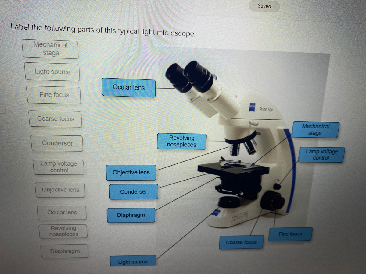
Answered: Saved Label the following parts of this… | bartleby
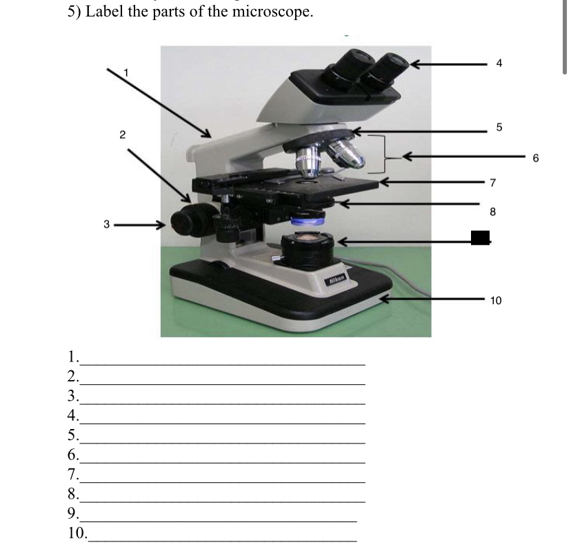
Answered: 5) Label the parts of the microscope. 1… | bartleby

Meiji MT6500 Series PCM NIOSH 7400 Asbestos Microscope
Labeling the Parts of the Microscope | Microscope World Resources

Microscope labeling
Microscope- Simple-AND Compound-WITH- Label - BS in Education ...

Collection Of Free Microscopes Drawing Label Clipart ...

Cytology - BIO 1210: Human Anatomy and Physiology I ...

Buy Levenhuk 500T Trinocular Microscope in online shop ...

Parts of a microscope with functions and labeled diagram

Lable the microscope worksheet

Photo Compound microscope with labels Image #3850568

Compound Microscope Parts, Functions, and Labeled Diagram ...

B3-220PL Educational Binocular Microscope | Motic Microscopes
Microscope with labels picture

Biology label part of microscope

Label the light microscope | Teaching Resources
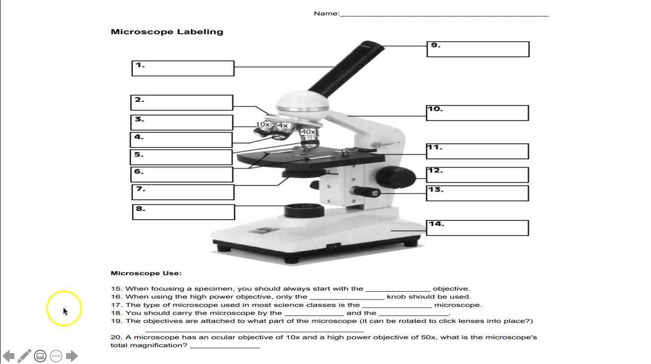
Parts of the Microscope Labeling Activity!
Post a Comment for "44 microscope images with labels"